Vada Spurgin
  • Blog
  • About
  • Contact

Should I Care For Calcaneal Apophysitis From Home?

5/18/2015

 
Overview

Sever?s disease, also known as calcaneal apophysitis or Osgood-Schlatter syndrome of the foot. This traction apophysitis is secondary to repetitive microtraumata or overuse of the heel in young athletes. The calcaneus is situated at the most plantar posterior aspect of the foot. The Achilles tendon inserts to the lower, posterior and slightly medial aspect of the calcaneus. The plantar fascia originates from the medial tubercle on the plantar aspect of the calcaneus. Proximal to the epiphysis is the apophysis, where the Achilles tendon actually inserts. The calcaneal growth plate and apophysis are situated in an area subject to high stress from the plantar and Achilles tendon.

Causes

Sever?s disease is caused by repetitive tension and/or pressure on the growth center of the heel. Running and jumping place a large amount of pressure on the heels and can cause pain. Children with Sever?s may limp or have an altered gait due to the pain. Risk factors for Sever's include tight calf muscles, weak ankle muscles, and alignment abnormalities at the foot and ankle. Sever?s can also result from wearing shoes without sufficient heel padding or arch support.

Symptoms

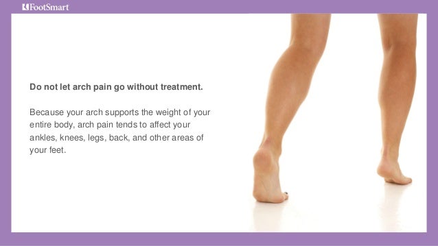
Unilateral or bilateral heel pain. Heel pain during physical exercise, especially activities that require running or jumping or are high impact. Pain is often worse after exercise. A tender swelling or bulge on the heel that is painful on touch. Limping. Calf muscle stiffness first thing in the morning.

Diagnosis

Sever?s disease can be diagnosed based on the symptoms your child has. Your child?s doctor will conduct a physical examination by squeezing different parts of your child?s foot to see if they cause any pain. An X-ray may be used to rule out other problems, such as a broken bone or fracture.

Non Surgical Treatment

The doctor might recommend that a child with Sever's disease. perform foot and leg exercises to stretch and strengthen the leg muscles and tendons, elevate and apply ice (wrapped in a towel, not applied directly to the skin) to the injured heel for 20 minutes two or three times per day, even on days when the pain is not that bad, to help reduce swelling, use an elastic wrap or compression stocking that is designed to help decrease pain and swelling, take an over-the-counter medicine to reduce pain and swelling, such as acetaminophen (Tylenol) or ibuprofen (Advil, Motrin).

Pain In The Foot's Arch What Are The Triggers ?

4/15/2015

 
Overview

Flat feet and fallen arches are terms used to describe lowering of the long inner arch of the foot. In the past, we thought that flat feet were a sign of a poorly developed or poorly structured foot. Now we know that people with flat feet function generally well and that flat feet don?t cause many foot problems. The most important factor in foot soreness and injury is not how flat or high your arches are, but the way you walk and move. If your feet move abnormally while you are walking or standing, this can make you more prone to injuries and foot soreness.

Arch Pain

Causes

Flat feet are a common condition. The condition is normal in infants and toddlers. Flat feet occur because the tissues holding the joints in the foot together (called tendons) are loose. The tissues tighten and form an arch as children grow older. This will take place by the time the child is 2 or 3 years old. Most people have normal arches by the time they are adults. However, the arch may never form in some people. Aging, injuries, or illness may harm the tendons and cause flat feet to develop in a person who has already formed arches. This type of flat foot may occur only on one side. Rarely, painful flat feet in children may be caused by a condition in which two or more of the bones in the foot grow or fuse together. This condition is called tarsal coalition.

Symptoms

Plantar fasciitis is most often seen in middle-aged men and women, but can be found in all age groups. The condition is diagnosed with the classic symptoms of pain well focused deep in the heel area of the bottom of the foot. Often the pain from plantar fasciitis is most severe when you first stand on your feet in the morning. Pain often subsides quite quickly, but then returns after prolonged standing or walking. Plantar fasciitis is sometimes, but not always, associated with a rapid gain of weight. It is also sometimes seen in recreational athletes, especially runners. In these athletes, it is thought that the repetitive nature of the sports causes the damage to the fibrous tissue that forms the arch of the foot.

Diagnosis

A patient is asked to step with full body weight on the symptomatic foot, keeping the unaffected foot off the ground. The patient is then instructed to "raise up on the tip toes" of the affected foot. If the posterior tibial tendon has been attenuated or ruptured, the patient will be unable to lift the heel off the floor and rise onto the toes. In less severe cases, the patient will be able to rise on the toes, but the heel will not be noted to invert as it normally does when we rise onto the toes. X-rays can be helpful but are not diagnostic of the adult acquired flatfoot. Both feet, the symptomatic and asymptomatic - will demonstrate a flatfoot deformity on x-ray. Careful observation may show a greater severity of deformity on the affected side.

Non Surgical Treatment

Flat feet in a child do not need treatment if they are not causing pain or walking problems. Your child's feet will grow and develop the same, whether special shoes, shoe inserts, heel cups, or wedges are used. Your child may walk barefoot, run or jump, or do any other activity without making the flat feet worse. In older children and adults, flexible flat feet that do not cause pain or walking problems do not need further treatment. If you have pain due to flexible flat feet, the following may help. An arch-support (orthotic) that you put in your shoe. You can buy this at the store or have it custom-made. Special shoes. Rigid or painful flat feet need to be checked by a health care provider. The treatment depends on the cause of the flat feet. For tarsal coalition, treatment starts with rest and possibly a cast. Surgery may be needed if pain does not improve. In more severe cases, surgery may be needed to clean or repair the tendon, fuse joints in the foot into a corrected position. Flat feet in older adults can be treated with pain relievers, orthotics, and sometimes surgery.

Arch Pain

Surgical Treatment

The procedure involves cutting and shifting the bone, and then performing a tendon transfer. First, the surgeon performs a calcaneal osteotomy, cutting the heel bone and shifting it into the correct position. Second, the surgeon transfers the tendon. Reroute the flexor digitorum to replace the troublesome posterior tibial tendon. Finally, the surgeon typically performs one or more fine-tuning procedures that address the patient?s specific foot deformity. Often, the surgeon will lengthen the Achilles tendon because it is common for the mispositioned foot to cause the Achilles to tighten. Occasionally, to increase the arch, the surgeon performs another osteotomy of one of the bones of the midfoot. Occasionally, to point the foot in a straightforward direction, the surgeon performs another osteotomy of the outside portion of the calcaneus.

Prevention

Stretch and strengthen important muscles in your feet, ankles and legs in order to guard against future strain. Make sure to acquire suitable arch supports and inserts if necessary, and that your shoes are shock absorbent and in good condition. Wearing tattered shoes provides no protection, and runners should replace their footwear before exceeding 500 miles of usage. Athletes new to arch supports should gradually build their training routine, allowing their feet to become accustomed to a new stance.

Stretching Exercises

Flexibility is important in preventing injuries. With a simple stretching exercise, you can rehabilitate the muscles of your foot to relieve arch pain and prevent future injuries. This simple exercise by Tammy White and Phyllis Clapis for Relay Health is a good way to strengthen your foot muscles and stretch your plantar fascia. Sit in a chair and cross one foot over your other knee. Grab the base of your toes and pull them back toward your leg until you feel a comfortable stretch. Hold 15 seconds and repeat three times. When you can stand comfortably on your injured foot, you can begin standing to stretch the plantar fascia at the bottom of your foot.

The Treatment And Cause Of Achilles Tendon Pain

4/3/2015

 
Overview

Achilles TendonAchilles Tendinitis is a common overuse injury which results in inflammation of the achilles tendon, most frequently causing mild to severe heel pain. The achilles tendon is the largest tendon in the body, connecting your calf muscle to your heel bone. It?s used with every step - when you walk, run, and jump. Although the Achilles tendon can withstand great stresses, it?s also prone to tendinitis. The condition is very common in athletes, especially runners who?ve suddenly increased the intensity or duration of their workouts. It?s also common in middle-aged, ?weekend athletes? who play sports like tennis or basketball only occasionally. The pain from achilles tendinitis may be felt anywhere from the back of the leg to the top of the heel. Most cases are mild and can be treated at home under a podiatrist?s supervision. Severe cases of Achilles tendinitis can lead to tendon tears (ruptures) that may require surgical repair.

Causes

Achilles tendonitis is aggravated by activities that repeatedly stress the tendon, causing inflammation. In some cases even prolonged periods of standing can cause symptoms. In many people who have developed achilles tendonitis, chronic shortening of the gastroc-soleus muscle complex is the reason that home remedies and anti-inflammatory medications fail. In these instances the muscle itself becomes shortened and creates a constant stress at the tendon?s attachment. Like a green branch that is slowly bent, eventually it begins to breakdown. Over a prolonged period the tendon becomes inflamed, and in the worst cases, appears swollen and thickened. In certain circumstances attempts to heal have failed and the body?s inability to heal the tissue results in degenerative changes known as achilles tendonosis. Anti-inflammatory medication, stretching and ice may only provide temporary relief, because they address the inflammation but not the root cause.

Symptoms

Achilles tendonitis typically starts off as a dull stiffness in the tendon, which gradually goes away as the area gets warmed up. It may get worse with faster running, uphill running, or when wearing spikes and other low-heeled running shoes. If you continue to train on it, the tendon will hurt more sharply and more often, eventually impeding your ability even to jog lightly. About two-thirds of Achilles tendonitis cases occur at the ?midpoint? of the tendon, a few inches above the heel. The rest are mostly cases of ?insertional? Achilles tendonitis, which occurs within an inch or so of the heelbone. Insertional Achilles tendonitis tends to be more difficult to get rid of, often because the bursa, a small fluid-filled sac right behind the tendon, can become irritated as well.

Diagnosis

If Achilles tendonitis is suspected, avoid any exercise or activity that causes the pain. It is advisable to see a doctor promptly so that an accurate diagnosis can be made and appropriate treatment recommended. The doctor will take a full medical history and will ask about the nature and duration of the symptoms. They will perform a physical examination of the affected area. Ultrasound scanning may be used to assess damage to the tendon or surrounding structures. Occasionally MRI (magnetic resonance imaging) may be recommended. The symptoms of Achilles tendonitis are often similar to symptoms of other conditions such as partial Achilles tendon rupture and heel bursitis. This can make diagnosis difficult and a referral to an orthopaedic specialist may be required in order for an accurate diagnosis to be made.

Nonsurgical Treatment

Most of the time, treatment for achilles tendinitis beginning with nonsurgical options. Your CFO physician may recommend rest, ice, ibuprofen, and physical therapy. If after 6 months, the pain does not improve, surgical treatment may be necessary. The type of surgery would depend on the exact location of the tendinitis and extent of damage.

Achilles Tendon

Surgical Treatment

If non-surgical treatment fails to cure the condition then surgery can be considered. This is more likely to be the case if the pain has been present for six months or more. The nature of the surgery depends if you have insertional, or non-insertional disease. In non-insertional tendonosis the damaged tendon is thinned and cleaned. The damage is then repaired. If there is extensive damage one of the tendons which moves your big toe (the flexor hallucis longus) may be used to reinforce the damaged Achilles tendon. In insertional tendonosis there is often rubbing of the tendon by a prominent part of the heel bone. This bone is removed. In removing the bone the attachment of the tendon to the bone may be weakened. In these cases the attachment of the tendon to the bone may need to be reinforced with sutures and bone anchors.

Prevention

Warm up slowly by running at least one minute per mile slower than your usual pace for the first mile. Running backwards during your first mile is also a very effective way to warm up the Achilles, because doing so produces a gentle eccentric load that acts to strengthen the tendon. Runners should also avoid making sudden changes in mileage, and they should be particularly careful when wearing racing flats, as these shoes produce very rapid rates of pronation that increase the risk of Achilles tendon injury. If you have a tendency to be stiff, spend extra time stretching. If you?re overly flexible, perform eccentric load exercises preventively. Lastly, it is always important to control biomechanical alignment issues, either with proper running shoes and if necessary, stock or custom orthotics.

What Causes Achilles Tendon Pain ?

4/2/2015

 
Overview

Achilles TendinitisAchilles tendinitis is an irritation/inflammation in the achilles tendon, which attaches to the back of the heel. It is often a result of overuse and occurs frequently in runners who have altered their training suddenly, either with regard to duration or intensity. This injury is also prevalent in middle-aged people who are active.
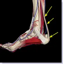
Causes

There are hundreds of tendons scattered throughout our body, but it tends to be a small handful of specific tendons that cause problems. These tendons usually have an area of poor blood supply that leads to tissue damage and poor healing response. This area of a tendon that is prone to injury is called a "watershed zone," an area when the blood supply to the tendon is weakest. In these watershed zones, they body has a hard time delivering oxygen and nutrients necessary for tendon healing, that's why we see common tendon problems in the same parts of the body. Tendonitis is most often an overuse injury. Often people begin a new activity or exercise that causes the tendon to become irritated. Tendon problems are most common in the 40-60 year old age range. Tendons are not as elastic and forgiving as in younger individuals, yet bodies are still exerting with the same force. Occasionally, there is an anatomical cause for tendonitis. If the tendon does not have a smooth path to glide along, it will be more likely to become irritated and inflamed. In these unusual situations, surgical treatment may be necessary to realign the tendon.

Symptoms

Achilles tendonitis is an injury that occurs when your Achilles tendon -- the large band of tissues connecting the muscles in the back of your lower leg to your heel bone -- becomes inflamed or irritated. The signs and symptoms of Achilles tendonitis often develop gradually. You'll feel pain and stiffness in your Achilles, especially when you first get out of bed. The pain lessens as you warm up, and may even disappear as you continue running. Once you stop, the pain returns and may feel even worse. You may also notice a crackling or creaking sound when you touch or move your Achilles tendon.

Diagnosis

The doctor will perform a physical exam. The doctor will look for tenderness along the tendon and pain in the area of the tendon when you stand on your toes. X-rays can help diagnose bone problems. An MRI scan may be done if your doctor is thinking about surgery or is worried about the tear in the Achilles tendon.

Nonsurgical Treatment

Make sure that the tendon is not torn through and through. If it is severed, you must see a doctor immediately so that the tendon can be repaired. Severe injuries can sever a tendon, without a skin laceration being present. Testing involves moving the toes and foot to see if the tendon moves. If the tendon does not appear to move, it may be severed (comparing the injured tendon and its movement to the same tendon on the uninjured foot may help). Very sharp pain, a sudden pop, or an obvious gap in the structure of the tendon are all signs of a rupture, and should be seen by a doctor as soon as possible. If there is extreme swelling of the leg, and pain (out of proportion to the amount of trauma received), you may have sustained a vascular injury. A doctor must see this type of injury immediately. If you are not sure, see a doctor. If you have multiple injured areas see a doctor immediately, in order to prevent excessive swelling and pain. If the above exam is negative, then you may proceed with self-treatment. (However, if you are not sure of the extent of your injury, you should consult your doctor immediately). The sooner you begin to treat your injury by following "R.I.C.E.", the better you will feel. Rest is very important. Take off your shoe, get off your feet, and relax. Ice should be applied as soon as possible. Never apply ice directly on the injured area, as the cold may make the pain worse. Ice should be applied close to the injured site, between the heart and the injury, so that as the blood flows under the ice, it will be cooled. This cool blood flowing into the injured area will help to reduce the swelling and pain. Apply the ice, wrapped in a cloth or over an elastic bandage, to the foot for 15 minutes, every 1-2 hours, for up to 3 days after an injury. If the ice is uncomfortable, or causes increased pain, do not continue to use it, and see a doctor. If you have poor circulation do not use ice, as this may cause a serious problem. c. Compression is used to limit swelling, and to give support to the injured area. Compression should be applied to the entire foot, starting first at the toes and working back to the ankle. If it is applied just to the injured area, increased swelling will occur in front and behind the wrapping. Compression should be applied with a 3-inch elastic bandage, beginning around the base of all the toes, and then going around the foot and ankle. Continue over the calf muscle when possible. Compression reduces motion in the injured area and foot, and this decreases the pain, and allows for quicker healing. The bandage should not be so tight that it causes increased pain or throbbing in the toes or foot. It should be comfortable! Do not remove the elastic bandage for the first 12 hours, unless it becomes too tight, or the pain increases, or the toes become pale, blue, or cool. If any of these things happen, immediately remove all bandages, and leave them off for several hours. The normal color and temperature of the toes should return immediately. If not, see a doctor immediately! Continue until the swelling and pain subsides; it could take from several days to several weeks. d. Elevation of the leg will aid in reducing swelling and pain. Blood rushes to an injured area to bring increased blood cells, that aid in healing. Gravity will also force blood to the injured area. Too many cells and too much fluid will apply pressure to the injured nerves and tissues, and cause increased pain and delayed healing. Keep your foot elevated so that it is at least parallel to the ground, or higher if it is comfortable. Do this for at least 48 hours, or until the throbbing subsides, when you lower the leg.

Achilles Tendinitis

Surgical Treatment

Around 1 in 4 people who have persisting pain due to Achilles tendinopathy has surgery to treat the condition. Most people have a good result from surgery and their pain is relieved. Surgery involves either of the following, removing nodules or adhesions (parts of the fibres of the tendon that have stuck together) that have developed within the damaged tendon. Making a lengthways cut in the tendon to help to stimulate and encourage tendon healing. Complications from surgery are not common but, if they do occur, can include problems with wound healing.

Prevention

There are several things you can do to reduce the risk of Achilles tendinitis, warm up every time before you exercise or play a sport. Switch up your exercises. Slowly increase the length and intensity of your workouts. Keep your muscles active and stay in shape all year-round. When you see symptoms of Achilles tendinitis, stop whatever activity you are doing and rest.

What Is Pes Planus?

3/31/2015

 
Overview

Acquired Flat Feet

When standing, most people have a gap under the arch of their foot. Flat feet, or pes planus is the term used to describe collapsed arches, meaning that the entire sole of the foot rests partially or completely on the ground. A simple way to check to see if you have flat feet is to wet your feet and step onto a dry, flat surface. Step away from your footprint; if you see a complete footprint, you more than likely have flat feet. If you only see toes, the pads of your feet, and your heels, you probably do not have flat feet. People who have flat feet usually roll their feet over to the inner side when they walk. The feet may even appear to point outward because of this movement. In most cases, the condition cannot be prevented.


Causes

Fallen arches in adults are caused by several things. Below are some of the most common causes. Abnormalities present from birth. Torn or stretched tendons (resulting from foot injuries or foot strains). Inflammation or damage of the PTT (posterior tibial tendon). The PTT is responsible for connecting the middle of the arch to the ankle and lower leg. Dislocated or broken bones (also as a result of injury). Health problems like rheumatoid arthritis. Nerve problems. Other factors like diabetes, obesity, aging and pregnancy (these factors are known to increase the risk of fallen arches).


Symptoms

A significant number of people with fallen arches (flat feet) experience no pain and have no problems. Some, however, may experience pain in their feet, especially when the connecting ligaments and muscles are strained. The leg joints may also be affected, resulting in pain. If the ankles turn inwards because of flat feet the most likely affected areas will be the feet, ankles and knees. Some people have flat feet because of a developmental fault during childhood, while others may find that the problem develops as they age, or after a pregnancy. There are some simple devices which may prevent the complications of flat feet.


Diagnosis

Diagnosis of flat feet or fallen arches can be made by your health practitioner and is based on the following. Clinical assessment involving visual gait assessment, as well as biomechanical assessment. A detailed family and medical history. A pain history assessment determining the location of painful symptoms. Physical palpation of the feet and painful areas. Imaging such as MRI or x-ray can be used by your practitioner to assist in the diagnosis.


Non Surgical Treatment

The treatment your podiatrist recommends will depend upon the trouble and pain you?re experiencing. Custom shoe inserts (orthotics) are most often recommended and are quite effective. Stretching exercises to loosen and strengthen the supporting tendons may also be recommended. Orthotic devices or bracing. To give your arch the support it needs, your foot and ankle surgeon may recommend an ankle brace or a custom orthotic device that fits into your shoe to support the arch. A short-leg cast or boot may be worn to immobilize the foot and allow the tendon to heal. Ultrasound therapy and stretching exercises may help rehabilitate the tendon and muscle following immobilization. Nonsteroidal anti-inflammatory drugs (NSAIDs), such as ibuprofen, help reduce the pain and inflammation. Your foot and ankle surgeon may recommend changes in your footwear.


Surgical Treatment

Flat Foot

Procedures may include the following. Fusing foot or ankle bones together (arthrodesis). Removing bones or bony growths, also called spurs (excision). Cutting or changing the shape of the bone (osteotomy). Cleaning the tendons' protective coverings (synovectomy). Adding tendon from other parts of your body to tendons in your foot to help balance the "pull" of the tendons and form an arch (tendon transfer). Grafting bone to your foot to make the arch rise more naturally (lateral column lengthening).

Learn How To Combat Pes Planus

3/31/2015

 
Overview

Acquired Flat Feet

Flat feet are a common problem. They can easily be identified by even non-trained individuals as the appearance is quite clear. On the inside of the foot should be a nice curvature which raises the inner foot up off the floor. If the foot touches the floor consistently from heel to toes then the foot is considered flat.


Causes

Most commonly fallen arches in adulthood can arise due to Posterior Tibial Tendon Dysfunction (PTTD). The posterior tibial tendon plays a pivotal role in holding up the arch of the foot, damage to this tendon results in the arch collapsing. Another cause of acquired flat feet in adulthood may be due to Rheumatoid arthritis as this disease attacks the bone, cartilage and ligaments in the foot causing it to change shape and flatten. Injury to the ligament, fracture and/or dislocation of the bones in the mid foot can all lead to a flat foot deformity. Adult acquired flat fleet can also arise in people who have diabetes or a neurological condition which limits normal feeling in the foot, the muscles in feet switch off, the tendons and ligaments weaken, and the arch eventually collapses.


Symptoms

A symptom is something the patient feels and reports, while a sign is something other people, including the doctor may detect. An example of a symptom may be pain in the ankle, while a sign may be a swelling. Symptoms may vary and generally depend on the severity of the condition. Some have an uneven distribution of bodyweight and find that the heel of their shoes wears out more rapidly and more on one side than the other. The most common signs or symptoms of flat feet are pain in the ankle (inner side), there may also be swelling of the foot in general, swelling in the arch of the foot, the calf, knee, the hip, the back, the general lower leg area. People with flat feet may also experience stiffness in one or both feet. One or both feet may be flat on the ground (either no arch, or very slight arch). Shoes may wear unevenly.


Diagnosis

It is important for people with foot pain to know if they have flat feet. The following tests can help you determine your arch type. When you get out of a swimming pool, look at your footprint on the concrete. The front of the foot will be joined to the heel by a strip. If your foot is flat, then the strip is the same width as the front of the foot, creating a footprint that looks like a stretched out pancake. With a normal arch, the strip is about half the width of the front of the foot. If you have a high arch, only a thin strip connects the front of the foot with the heel. Put your shoes on a flat table and view them at eye level from behind. See if the sole is worn evenly. A flat foot will cause more wear on the inside of the sole, especially in the heel area. The shoe will easily rock side to side. A flat foot will also cause the upper part of the shoe to lean inward over the sole. Both shoes should wear about the same way. If you have pain in one foot, you should make sure you don't have a fallen arch on that side. There are two good tests you can perform at home to detect this problem. Place your fingertips on a wall that you are directly facing and stand on your tiptoes on one foot. If you can't do it, a fallen arch may be the culprit. Stand with your feet parallel. Have someone stand in back of you and look at your feet from behind. You can also do it yourself if you stand with your back to a mirror. Normally, only the pinky toe is visible from behind. If one foot is flatter than the other, the 4th and sometimes the 3rd toe on that foot can also be seen.


Non Surgical Treatment

Get shoes made for walking or running. One way to support your arch is to wear good-quality running or walking shoes, says Dr. Gastwirth. "These shoes generally provide good support to the foot." Add support. The top-of-the-line arch support is an orthotic insole, which may cost $900 or more and must be custom-made. "But many people with sore arches will get relief with over-the-counter arch supports for about $10," suggests Judith Smith, M.D., assistant professor of orthopedic surgery at Emory University School of Medicine in Atlanta. "The thing to remember about arch supports is that your shoe must have enough depth to accommodate them. Otherwise, you'll get a lot of rubbing on the top of your foot, or your heel will come out of the shoe." Most mens shoes are deep enough to accommodate the insoles; women should take their shoes with them to the drugstore when buying the insoles to ensure a good fit. If your heels are high, keep them wide. High heels may be your Achilles' heel--especially if you wear them constantly. "Flatter shoes are no doubt better," says Dr. Sanfilippo. Flat heels help prevent fallen arches and are kinder to your feet if fallen arches have already occurred. "If you must wear high heels, choose styles with a wide heel. Stay away from stiletto heels."


Surgical Treatment

Flat Foot

Surgery for flat feet is separated into three kinds: soft tissue procedures, bone cuts, and bone fusions. Depending on the severity of the flat foot, a person?s age, and whether or not the foot is stiff determines just how the foot can be fixed. In most cases a combination of procedures are performed. With flexible flat feet, surgery is geared at maintaining the motion of the foot and recreating the arch. Commonly this may involve tendon repairs along the inside of the foot to reinforce the main tendon that lifts the arch. When the bone collapse is significant, bone procedures are included to physically rebuild the arch, and realign the heel. The presence of bunions with flat feet is often contributing to the collapse and in most situations requires correction. With rigid flat feet, surgery is focused on restoring the shape of the foot through procedures that eliminate motion. In this case, motion does not exist pre-operatively, so realigning the foot is of utmost importance. The exception, are rigid flat feet due to tarsal coalition (fused segment of bone) in the back of the foot where freeing the blockage can restore function.


Prevention

Strap the arches into the anatomically correct positions with athletic tape and leave them like this for some time. If the fallen arches are an issue with the muscular structure, this may give the muscles an opportunity to strengthen. This is definitely not a fallen arches cure all the time but it can help prevent it more times than not. Ask a doctor or physical therapists to show you how to do this taping. Find shoes that fit. This may require that you get your foot measured and molded to ensure that the shoe will fit. Shoes that are too big, too tight or too short, may not directly cause the fallen arches, but they can assist with the damage to the area. These shoes should have thick cushioning inside and have plenty of room for your toes. Walk without shoes as much as possible. Shoes directly assist with weakening and distorting the arches of the feet so going without shoes can actually help strengthen your arches and prevent fallen arches. Walking on hard and bumpy surfaces barefooted makes the muscles in your feet strengthen in order to prevent injury. It is a coping mechanism by your body. Insert heel cups or insoles into the shoes that you wear the most. Many people wear uncomfortable shoes to work and these are the same shoes that cause their arches the most problems. Inserting the heel cups and insoles into these shoes can prevent fallen arches from occurring. Many people place these inserts into all their shoes to ensure support. Ask a medical professional, either your doctor or a physical therapist, about daily foot exercises that may keep the arches stronger than normal. Many times, you can find exercises and stretches on the Internet on various websites. Curling your toes tightly and rotating your feet will help strengthen your longitudinal arches. Relax your feet and shake them for a minute or so before you do any arch exercises. This will loosen the muscles in your feet that stay tight due to normal daily activities. Wear rigid soled sandals whenever possible to provide a strong support for your arches. Wooden soled sandals are the best ones if available. Walk or jog on concrete as much as you can. This will create a sturdy support for your arches. Running or walking in sandy areas or even on a treadmill, does not give rigid support. Instead, these surfaces absorb the step, offering no support for arches.


After Care

Patients may go home the day of surgery or they may require an overnight hospital stay. The leg will be placed in a splint or cast and should be kept elevated for the first two weeks. At that point, sutures are removed. A new cast or a removable boot is then placed. It is important that patients do not put any weight on the corrected foot for six to eight weeks following the operation. Patients may begin bearing weight at eight weeks and usually progress to full weightbearing by 10 to 12 weeks. For some patients, weightbearing requires additional time. After 12 weeks, patients commonly can transition to wearing a shoe. Inserts and ankle braces are often used. Physical therapy may be recommended. There are complications that relate to surgery in general. These include the risks associated with anesthesia, infection, damage to nerves and blood vessels, and bleeding or blood clots. Complications following flatfoot surgery may include wound breakdown or non-union (incomplete healing of the bones). These complications often can be prevented with proper wound care and rehabilitation. Occasionally, patients may notice some discomfort due to prominent hardware. Removal of hardware can be done at a later time if this is an issue. The overall complication rates for flatfoot surgery are low.

Does Pain In The Arch Require Surgical Procedures ?

3/30/2015

 
Overview

Most flat feet are not painful, particularly those flat feet seen in children. In the adult acquired flatfoot, pain occurs because soft tissues (tendons and ligaments) have been torn. The deformity progresses or worsens because once the vital ligaments and posterior tibial tendon are lost, nothing can take their place to hold up the arch of the foot. The painful, progressive adult acquired flatfoot affects women four times as frequently as men. It occurs in middle to older age people with a mean age of 60 years. Most people who develop the condition already have flat feet. A change occurs in one foot where the arch begins to flatten more than before, with pain and swelling developing on the inside of the ankle. Why this event occurs in some people (female more than male) and only in one foot remains poorly understood. Contributing factors increasing the risk of adult acquired flatfoot are diabetes, hypertension, and obesity.

Arch Pain

Causes

There are many different factors that can cause arch pain. A structural imbalance or an injury to the foot can often be the direct cause. However, most frequently the cause is a common condition called plantar fasciitis. The plantar fascia is a broad band of fibrous tissue located along the bottom surface of the foot that runs from the heel to the forefoot. Excessive stretching of the plantar fascia, usually due to over-pronation (flat feet), causes plantar fasciitis. The inflammation caused by the plantar fascia being stretched away from the heel often leads to pain in the heel and arch areas. The pain is often extreme in the morning when an individual first gets out of bed or after a prolonged period of rest. If this condition is left untreated and strain on the longitudinal arch continues, a bony protrusion may develop, known as a heel spur. It is important to treat the condition promptly before it worsens.

Symptoms

The foot of a newborn with congenital vertical talus typically has a convex rocker-bottom shape. This is sometimes combined with an actual fold in the middle of the foot. The rare person who is diagnosed at an older age often has a "peg-leg" gait, poor balance and heavy calluses on the soles where the arch would normally be. If a child with congenital vertical talus has a genetic disorder, additional symptoms often are seen in other parts of the body.

Diagnosis

The adult acquired flatfoot, secondary to posterior tibial tendon dysfunction, is diagnosed in a number of ways with no single test proven to be totally reliable. The most accurate diagnosis is made by a skilled clinician utilizing observation and hands on evaluation of the foot and ankle. Observation of the foot in a walking examination is most reliable. The affected foot appears more pronated and deformed compared to the unaffected foot. Muscle testing will show a strength deficit. An easy test to perform in the office is the single foot raise.

Non Surgical Treatment

For mild pain or aching, acetaminophen (Tylenol) or a nonsteroidal anti-inflammatory drug (NSAID), such as aspirin or ibuprofen (Advil, Motrin and others) may be effective. When there are no symptoms, treatment is not needed. If a child older than age 3 develops symptoms, the doctor may prescribe a therapeutic shoe insert made from a mold of the child's foot or a corrective shoe. As an alternative, some doctors recommend store-bought arch supports. These appear to work as well as more expensive treatments in many children. With any conservative, nonsurgical treatment, the goal is to relieve pain by supporting the arch and correcting any imbalance in the mechanics of the foot. Surgery is typically offered as a last resort in people with significant pain that is resistant to other therapies.

Pain In Arch

Surgical Treatment

There are two types of bone procedure for flat feet, those where bone cuts and bone grafts are used to alter the alignment by avoiding any joint structures, or joint invasive procedures (called fusions or arthrodeses) that remove a joint to reshape the foot. With joint fusion procedures, there are those procedures that involve non-essential joints of the foot versus those that involve essential joints. All bone procedures have their place in flat foot surgery, and Dr. Blitz carefully evaluates each foot to preserve as much motion and function while obtaining proper and adequate alignment. In many cases a flat foot reconstruction involves both soft tissue procedures and bone procedures to rebuild and restore the arch. There are several joints in the arch of the foot that can collapse - and these joints are non-essential joints of the foot. This does not mean that they do not have a purpose, but rather become inefficient is providing a stable platform for function. As such, locking these non-essential non-functioning joints into place is commonly recommended. These joints are fused together with screws and/or plates. A heel bone that is no longer in proper position and pushed outwards away from the foot can be corrected with a bone cut and realignment procedure, so long as the displacement is not too significant. A benefit of this surgery is that it keeps the back portion of the foot mobile, and helps the surrounding tendons work for efficiently in maintaining the arch. In certain flat feet, the foot is deviated outwards and away from the midline of the body. Sometimes, this is due to the outer portion of the foot being shorter than the inner portion. Here bone graft can be added to the outer edge of the foot to lengthen the foot to swing the foot over into a corrected position. This procedure is most commonly performed in children and young adults. A bone graft is inserted into the top part of the arch to realign a component of the flat foot, medically known as forefoot varus or medial column elevatus. The back part of the foot (called the rearfoot complex) can be the cause (or source) of the flat foot or the simply affected by the flat foot foot. In simple terms, the back part of the foot can be made to flatten out due to arch problems - and vica versa for that matter. Dr. Blitz specifically identifies the cause of the flat foot as this will determine the best treatment plan, as each flat foot needs to be evaluated individually. The rearfoot is made up of three joints, and depending on the extent and most importantly the rigidity of these joints, they may require fusion to restore alignment. When all three joints require fusion - this call is a triple arthrodesis. For completeness, isolated fusion of any of the three joints can be performed (such as subtalar joint arthrodesis, talonavicular arthrodesis, and calcaneaocuboid joint arthrodesis). The medical decision making for isolated fusions is beyond the scope this article, but Dr. Blitz tries to avoid any rearfoot fusion for flexible feet because these are joints are essential joints of the foot, especially in younger people. Those in severe cases, it may be advantageous to provide re-alignment.

Prevention

Stretch and strengthen important muscles in your feet, ankles and legs in order to guard against future strain. Make sure to acquire suitable arch supports and inserts if necessary, and that your shoes are shock absorbent and in good condition. Wearing tattered shoes provides no protection, and runners should replace their footwear before exceeding 500 miles of usage. Athletes new to arch supports should gradually build their training routine, allowing their feet to become accustomed to a new stance.

Pain In The Arches Cause And Effect

3/29/2015

 
Overview

The arch of the foot is a collection of bones, ligaments, tendons, and muscles that are constructed to allow your foot to bear the weight of your body safely. These tissues and bones of your arch are in constant motion as you walk and run, absorbing impacts that might otherwise injure you. The arch of your foot has its limits, however, and arch pain can be a sign that you?ve passed those limits and injured yourself. Pay close attention to any pain you are feeling in your foot. What may seem at first to be simple soreness from being on your feet all day could be a sign of a more serious stress injury or repeated motion injury. Arch pain could also be a result of a fall or impact injury, so be sure to seek proper treatment.

Arch Pain

Causes

There are several reasons why arch pain develops. Sometimes it?s due to a condition known as plantar fasciitis, in which the plantar fascia (the band of tissue that runs along the bottom of your foot from your heel to your toes) becomes inflamed after excessive stress. Heel pain results from this inflammation. Sometimes the pain is due to extensive time spent on your feet. Many people feel pain on the arch of their feet after a long workday, while others overuse their feet exercising or playing sports. A foot deformity, such as hammertoe or clubfoot, can also cause this pain. Medical conditions such as diabetes or obesity can put additional stress on your feet, thereby causing arch pain. Your footwear is also important. Shoes should support all parts of your foot, especially the bottom. This is very important if you spend excessive time on your feet, if your obese, if your pregnant, or if you engage in sport-related activities. Injuries to any of the twenty-six bones, thirty-three joints and over 100 muscles, tendons and ligaments in the feet can also cause arch pain. Because the foot is such a complex structure, it?s important to see a podiatrist at the first sign of symptoms.

Symptoms

Pain in arch of foot is really the only symptom of this condition. It is unlikely to see any swelling or bruising and instead there will be a deep tender spot near the heel. Occasionally the pain may radiate further down the foot. With this condition, pain will usually be felt first thing in the morning or after periods of sitting. This is because the plantar fascia tightens and shortens slightly when there is no weight on it and by standing on it it suddenly stretches and becomes painful. After a few steps it starts to loosen off and the pain may subside. If this is the same pattern of pain you experience it is quite likely you have plantar fasciits. Pain may also be felt when walking up stairs or standing on tip-toes (anything that stretches the fascia).

Diagnosis

In people with flat feet, the instep of the foot comes in contact with the ground when standing. To diagnose the problem, the health care provider will ask you to stand on your toes. If an arch forms,the flat foot is called flexible. You will not need any more tests or treatment. If the arch does not form with toe-standing (called rigid flat feet), or if there is pain, other tests may be needed, including a CT scan to look at the bones in the foot. MRI scan to look at the tendons in the foot. X-ray of the foot.

Non Surgical Treatment

The treatment of a rigid flatfoot depends on its cause. Congenital vertical talus. Your doctor may suggest a trial of serial casting. The foot is placed in a cast and the cast is changed frequently to reposition the foot gradually. However, this generally has a low success rate. Most people ultimately need surgery to correct the problem. Tarsal coalition. Treatment depends on your age, extent of bone fusion and severity of symptoms. For milder cases, your doctor may recommend nonsurgical treatment with shoe inserts, wrapping of the foot with supportive straps or temporarily immobilizing the foot in a cast. For more severe cases, surgery is necessary to relieve pain and improve the flexibility of the foot. Lateral subtalar dislocation. The goal is to move the dislocated bone back into place as soon as possible. If there is no open wound, the doctor may push the bone back into proper alignment without making an incision. Anesthesia is usually given before this treatment. Once this is accomplished, a short leg cast must be worn for about four weeks to help stabilize the joint permanently. About 15% to 20% of people with lateral subtalar dislocation must be treated with surgery to reposition the dislocated bone.

Arch Pain

Surgical Treatment

Surgery may be necessary in situations where the symptoms are likely to get worse over time, or when pain and instability cannot be corrected with external orthopedic devices. There are many types of surgical procedures, including cavus foot reconstruction, which can be performed to correct the foot and the ankle and restore function and muscle balance.

Prevention

To prevent arch pain, it is important to build up slowly to your exercise routine while wearing arch supports inside training shoes. By undertaking these simple measures you can prevent the discomfort of arch pain which can otherwise linger for many months. While you allow the foot to recover, it will help to undertake low impact exercises (such as swimming or water aerobics).

Adult Aquired FlatFoot

3/27/2015

 
Overview

Adult acquired flatfoot can be a progressive disorder which involves the compromise involving soft tissue supports in the medial arch. the situation mostly affects middle outdated females and it is seen as a lowering in the arch, turning out in the forefoot, and a sideways angulation of the heel. You can easily find five stages in the disorder that will gets progressively disabling. the end stage could potentially compromise your ankle joint together with all the joints in the hindfoot.

Adult Acquired Flat Feet

Causes

Women are affected through Adult Acquired Flatfoot 4 times more frequently when compared with men. Adult Flatfoot typically occurs in center in order to older get older people. The Majority Of individuals who acquire the situation curently have flat feet. one arch begins to flatten more, then pain as well as swelling develop on the inside in the ankle. This situation generally affects just one foot. The idea is unclear why ladies are generally affected more often than men. However elements that may increase your risk of Adult Flatfoot include diabetes, hypertension, and also obesity.

Symptoms

Symptoms are minor and might even go unnoticed, Pain dominates, as opposed to deformity. Minor swelling could become noticeable across the length of the particular tendon. Pain as well as swelling over the course of the tendon. Noticeable decline in arch height. Aduction with the forefoot in rearfoot. Subluxed tali as well as navicular joints. Deformation at this point is even now flexible. Considerable deformity along with weakness. Significant pain. Arthritic changes inside the tarsal joints. Deformation as associated with this point is actually rigid.

Diagnosis

Starting in the knee down, examine for any bowing in the tibia. The tibial varum will trigger increased medial strain around the foot and ankle. This may be important to contemplate throughout surgical planning. Verify the actual gastrocnemius muscle mass along with Achilles complex by method of a new straight as well as bent knee examine for equinus. When the particular selection of motion improves to a minimal of neutral with bent knee testing of the Achilles complex, one may think regarding a gastrocnemius recession. If the particular Achilles complex is actually nevertheless tight with bent knee testing, an Achilles lengthening could end up being necessary. check the particular posterior tibial muscle alongside its entire course. Palpate the actual muscle as well as observe the tendon pertaining to power with a plantarflexion and inversion tension test. check the particular flexor muscles with regard to strength to become able to discover if a sufficient transfer tendon will be available. check your anterior tibial tendon pertaining to dimension and strength.

Non surgical Treatment

Because in the progressive nature associated with PTTD, early treatment method is actually advised. In the event that handled early enough, your signs and symptoms may resolve without having the requirement for surgery along with progression of your issue may be arrested. Throughout contrast, untreated PTTD could depart anyone having an extremely flat foot, painful arthritis inside the foot as well as ankle, as well as growing limitations about walking, running, or other activities. Throughout many cases regarding PTTD, treatment may start out with non-surgical approaches in which may include. Orthotic devices or bracing. To Always Be Able To give your own arch your assistance it needs, your current foot and also ankle surgeon could supply you by getting an ankle brace or possibly a custom orthotic device that fits in in order to the shoe. Immobilization. Sometimes a short-leg cast or even boot will be worn to immobilize the actual foot and permit the particular tendon to heal, or perhaps a person could have to entirely stay away from all weight-bearing for a while. Physical therapy. Ultrasound therapy as well as exercises could aid rehabilitate the tendon as well as muscle mass following immobilization. Medications. Nonsteroidal anti-inflammatory drugs (NSAIDs), for example ibuprofen, assist decrease the anguish and also inflammation. Shoe modifications. your foot and ankle surgeon could advise changes to create together together with your shoes and could supply unique inserts designed to boost arch support.

Adult Acquired Flat Foot

Surgical Treatment

For patients having a more severe deformity, or even significant signs as well as symptoms that do not react to conservative treatment, surgery may be necessary. You will find a number of procedures available depending about the naturel of one's condition. Ligament as well as muscle mass lengthening, removal of inflamed tendon lining, transferring of the close by tendon for you to re-establish an arch, and also bone realignment along with fusion are examples associated with surgical choices to assist having a painful flatfoot condition. Surgery could be avoided when symptoms are usually addressed early. In your event that you're sensation ankle pain or perhaps notice just about any warmth, redness as well as swelling within your foot, contact us immediately. We could create a tailored treatment method program to solve your signs and symptoms along with prevent long term problems.

The Causes And Also Treatment

3/26/2015

 
Overview

There are usually a few some other items that can weaken your tendon (and thus move in which quitting occasion a little closer). Females are generally far more most likely as compared to men for you to develop this condition, also it may take location across the exact same moment as menopause (around age 60 as well as so). Steroid use (not often illegal-this might originate from having cortisone shots inside the area) and smoking could also improve the chance with regard to developing PTTD, since steroids tend to weaken tendons. The history of injury within the area, arthritis, or even an already flat foot might also actually push the particular tendon to always be able to declare, ?That?s the final straw!? (Silly tendon. Because if it even understands what straw is.)

Acquired Flat Foot

Causes

There are generally multiple elements contributing for the development of this problem. Damage towards the nerves, ligaments, and/or tendons in the foot could cause subluxation (partial dislocation) in the subtalar as well as talonavicular joints. Bone fracture is a possible cause. the resulting joint deformity from any of these issues can lead to adult-acquired flatfoot deformity. Dysfunction with the posterior tibial tendon offers often been linked along with adult-acquired flatfoot deformity (AAFD). The Actual loss of active and also passive pull with the tendon alters the particular typical biomechanics with the foot and ankle. The Particular causes of this can end up being many along with varied as well. Diabetes, high blood pressure, and also prolonged use regarding steroids tend to be some involving the more common causes of adult-acquired flatfoot deformity (AAFD) brought upon by simply impairment in the posterior tibialis tendon. Overstretching or even rupture of the tendon brings about tendon as well as muscle mass imbalance inside the foot leading to adult-acquired flatfoot deformity (AAFD). Rheumatoid arthritis can be one of the greater widespread causes. Regarding 50 % of just about all adults with this particular kind involving arthritis will develop adult flatfoot deformity over time. Within such cases, the particular situation will be gradual along with progressive. Weight Problems has been linked with this particular condition. Loss regarding blood supply for practically any cause within the location in the posterior tibialis tendon is an additional factor. Various Other possible leads to contain bone fracture as well as dislocation, a new torn or perhaps stretched tendon, or even a neurologic condition causing weakness.

Symptoms

Pain as well as swelling at the particular rear of the within of your ankle and also together your current instep. An Individual might always be tender powering the particular inner ankle where the posterior tibial tendon programs and also sometimes get burning, shooting, tingling or perhaps stabbing pain as a consequence of inflammation with the nerve inside the particular tarsal tunnel. Problems walking, the particular inability simply for you to walk long mileage plus a generalised ache whilst strolling even short distances. This specific may most likely turn out for you to be more pronounced following each day. Adjust in foot shape, sometimes your current tendon stretches out, this truly is thanks to weakening in the tendon as well as ligaments. When this occurs, the arch in your foot flattens along using a flatfoot deformity occurs, presenting a new change in foot shape. Inability to tip-toe, any way of diagnosing Posterior Tibial Tendon Dysfunction can be trouble or perhaps inability to end up being able to ?heel rise? (stand on your toes using one foot). Your Current tibialis posterior tendon enables you for you to definitely perform this manoeuvre effectively. A Person might also encounter pain upon attempting to do the heel rise.

Diagnosis

Although you'll end up being in a new position to do the "wet test" at home, a comprehensive examination by a physician is going to be necessary to recognize why your flatfoot developed. Feasible leads to incorporate a congenital abnormality, a bone fracture or perhaps dislocation, a torn or stretched tendon, arthritis or even neurologic weakness. for example, an inability to rise on your toes whilst standing on the affected foot could indicate damage to the posterior tibial tendon (PTT), which supports the heel as well as types your arch. In the particular event that "too many toes" show around the outside of one's foot if the physician views anyone from the rear, your own shinbone (tibia) might end up being sliding off your anklebone (talus), an additional indicator of harm to the PTT. Always Be positive in order to put on your own standard shoes towards the examination. An irregular put on pattern on the bottom in the shoe is yet another indicator of acquired adult flatfoot. your physician may request X-rays to observe how the particular bones of your feet tend to be aligned. muscle and also tendon power are usually tested simply by asking to move your foot even although the doctor holds it.

Non surgical Treatment

Nonoperative remedy regarding posterior tibial tendon dysfunction may be successful with just about all the Arizona AFO brace, specially when treatment method will be initiated inside the first stages with the disease. This kind of mandates the orthopedist has a high index involving suspicion when evaluating patients to create a precise diagnosis. Though there's a role pertaining to surgical management regarding acquired flat feet, a new well-fitted, custom-molded leather along with polypropylene orthosis could be successful in relieving symptoms and also either obviating or perhaps delaying virtually any surgical intervention. Inside today's climate regarding patient fulfillment directed well being care, a new less invasive treatment modality in which relieves pain might prove to become more valuable when compared with similar pain relief that is obtained following surgery. Concerns relating to the long-term outcomes involving bracing remain unanswered. future studies tend to be required to see whether disease progression and also arthrosis occur despite symptomatic relief with a brace. Furthermore, age- along with disease stage-matched control teams who're randomized for you to undergo surgery or bracing are generally required to compare these different therapy modalities.

Flat Foot

Surgical Treatment

In your event that conservative treatment fails surgical intervention is actually offered. for any Stage 1 deformity the posterior tibial tendon tenosynovectomy (debridement of the tendon) or primary repair may always be indicated. Regarding Stage two any combination of Achilles lengthening along with bone cuts, calcaneal osteotomies, as well as tendon transfers is common. Stage 2 versatile PTTD may always be the most frequent stage patients existing together with pertaining to treatment. Within Stage three as well as 4 PTTD isolated fusions, locking a pair of or perhaps more joints together, maybe indicated. Most therapy is dependent on the stage and also severity from presentation with almost all the goals and activity amount individual inside mind. treatment can be customized towards the individual affected person needs.
<<Previous
Forward>>

    Archives

    July 2017
    June 2017
    May 2017
    February 2016
    September 2015
    August 2015
    June 2015
    May 2015
    April 2015
    March 2015

    Categories

    All

    RSS Feed

Powered by Create your own unique website with customizable templates.